Translate

Wednesday 21 May 2014

Biochip

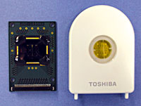
A biochip is a device that has some of the features of a computer chip but, instead of doing calculations, it uses living cells or molecules from living cells to greatly speed up certain laboratory tests. A typical biochip is a glass or plastic chip or tile a few inches on a side. It has hundreds or even tens of thousands of microscopic droplets of material stuck to its surface like gum on a sidewalk. A computer looks at the chip using a camera. Information from a biochip can be used to learn about the differences between genes, cells, or drugs. It can also be used to study many other questions about cells. Biochips are also called microarrays, where micro means ‘‘small’’ and an array is any regular grid, such as a chessboard. The droplets on a biochip are laid down in a checkerboard pattern. A square chip five inches on a side may have 40,000 or more spots on its surface.

The most common kind of biochip is the DNA microarray, also called a gene chip or DNA chip. Deoxyribonucleic acid (DNA) is the long, coded molecule used by all living things to pass on traits to offspring. DNA also tells each cell how to make all the molecules it needs to live, like a cookbook containing many recipes. The DNA of almost every living thing is at least slightly different from that of every other.

In one type of DNA chip, genes (short pieces of DNA that code for single molecules) are placed on the chip. Since even large molecules are too small to see with the naked eye, millions of copies of each gene can be placed on a tiny spot on the chip.


HOW DNA CHIP WORKS?


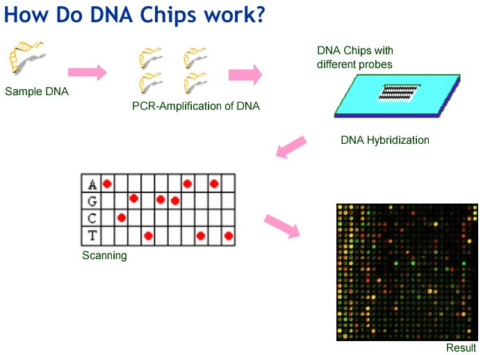
In a DNA chip, each separate spot (also called a probe) contains one type of defective gene. To find out if a person has any of these defective genes in their own DNA, DNA is taken from the person’s cells. Copies of the person’s DNA are made, and these copies are labeled, meaning that they include a chemical that glows when ultraviolet light (which is invisible to the eye) shines on it. Small drops of liquid containing labeled copies of the person’s DNA are then added to the spots on the biochip.

A normal DNA molecule is shaped like a ladder, but the DNA copies being mixed on the biochip are one-sided copies, like a ladder that has been sawed in half lengthwise, cutting every rung in half. When two pieces of one-sided DNA that have matching rungs (or bases, as they are called) meet, they lock or zip together. When this happens, the two pieces of DNA are said to hybridize. If the patient’s genes match any of the defective genes that have been put on the biochip, they will attach to (hybridize with) those defective genes.

The chip is then washed to remove any of the person’s DNA that has not found a match on the chip. Finally, the chip is placed in ultraviolet light, and a camera records any spots that glow. These are spots where the labeled copies of the person’s DNA have matched up with DNA on the chip.

Examining a patient’s DNA for defects is called genetic screening. By using a biochip, genetic screening can be done very quickly, all the tests can be done at once, rather than doing hundreds or even thousands of separate tests.

Genetic screening is only one way of using biochips. Another important use for biochips is to study how genes are used by living cells. Each gene tells the cell how to make a certain protein molecule. Cells read the recipe given by the gene by first making another molecule, mRNA, which copies the information in the gene. The mRNA can then go to a place in a cell that will build the molecule that the gene codes for. The more mRNA a cell has for a gene at a particular time, the more it is said to be ‘‘expressing’’ that gene, i.e, the more of that particular molecule it is making. Gene expression changes all the time for thousands of genes in every cell.

In the laboratory, scientists can make DNA molecules from the mRNA found in a cell. This matching DNA is called cDNA (complementary DNA). If a biochip has all the genes of an organism dotted on its surface, then cDNA made from the mRNA in a cell can attach to (hybridize with) the genes on the chip. The more a gene is being expressed in the cell, the more cDNA for that gene there will be, and the more that cDNA will stick to the matching genes on the biochip. Spots with more labeled cDNA will glow more brightly under ultraviolet light. In this way, scientists can literally take a snapshot of how the genes in a cell are being expressed at any one time—how much the cell is making, at that moment, of thousands of different substances. This is extremely useful in trying to understand how cancer cells grow and in many other medical problems.

How complex cells arose on earth?

Prokaryotes were the first life on earth. There are fossil bacteria that are 3.8 billion years old, nearly as old as the oceans themselves. Being prokaryotes, these early cells did not have organized organelles enclosed by membranes. Prokaryotic life came on the scene very early in the earth’s history, but the next step took much longer. It was a billion years or more before complex cells with organelles, or eukaryotic cells, appeared. Strangely enough, this advance in cell design, shared by all multicellular life, probably started with a simple meal.

Biologists believe that mitochondria, the “powerhouses” of the cell, were once free-living bacteria. They are 1 to 3 microns (μm) long, about the same size as many bacteria. Like many bacteria, they contain layers of folded membranes. Although most of the cell’s DNA is contained in the nucleus, mitochondria have a small amount of their own DNA. This DNA is even in the form of a single, circular molecule like the chromosome of bacteria. When a cell needs new mitochondria it does not make them from scratch. Instead, the mitochondria within the cell divide in two, much as bacteria divide by cell division. Mitochondria resemble bacteria in several other respects.


Because of these similarities, biologists believe that mitochondria were originally bacteria that came to live inside other cells. The most likely scenario is that relatively large prokaryotic cells that ate smaller ones were unable to digest all of their food. It is also possible that the small cells were disease bacteria that invaded the larger cell but were unable to kill it. Both phenomena have been observed in the laboratory in living cells. Either way, the small cells eventually took up permanent residence in the larger cells, using them as hosts. The living together of different kinds of organisms like this is called symbiosis.

According to this theory, the host cells could not use oxygen in respiration. The smaller, symbiotic, bacteria did use oxygen and gave this ability to their hosts. This gave the host cells an advantage over cells that didn't have symbiotic bacteria.

Over the course of time, the host cells were able to transfer most of the symbiotic bacteria’s genes into their own nuclei, and thus came to control the symbionts. The bacteria and host cells became more and more dependent on each other. Eventually the bacteria became mitochondria.


Chloroplasts, the organelles that perform photosynthesis, are also thought to have arisen from symbiotic bacteria. Like mitochondria, they are about the right size, have a circular DNA molecule, and can reproduce themselves. Chloroplasts also contain folded membranes very similar to those of photosynthetic bacteria that we see today. In fact, biologists have found a possible “missing link” between photosynthetic bacteria and chloroplasts. Some sea squirts and other invertebrates contain symbiotic, photosynthetic bacteria called Prochloron that are similar to the chloroplasts of green algae and higher plants. It is thought that these chloroplasts arose from Prochloron-like bacteria when host cells ate the bacteria, some of which managed to resist being digested and became symbiotic.

The hypothesis that mitochondria and chloroplasts arose from bacteria, once highly controversial, is now almost universally accepted by biologists. Some biologists now even classify mitochondria and chloroplasts as symbiotic bacteria rather than organelles. Several other types of organelles may have similar origins. All multicellular organisms, including humans, carry in their very cells the legacy of these bacterial partnerships, probably the result of an ancient snack.

Complex cell

Tuesday 20 May 2014

Use of Retroviral Vectors in Gene therapy

Some 24 per cent of all gene therapy clinical trials undertaken to date have employed retroviral vectors as gene delivery systems. Retroviruses are enveloped viruses. Their genome consists of ssRNA of approximately 8 kb. Upon entry into sensitive cells, the viral RNA is reverse transcribed and eventually yields double-stranded DNA. This subsequently integrates into the host cell genome. The basic retroviral genome contains a minimum of three structural genes : gag (codes for core viral protein), pol (codes for reverse transcriptase) and env (codes for the viral envelope proteins).

Retroviral life cycle


At either end of the viral genome are the long terminal repeats (LTRs), which harbour powerful promoter and enhancer regions and sequences required to promote integration into the host DNA. Also present, immediately adjacent to the 5' LTR, is the packing sequence (ψ). This is required to promote viral RNA packaging.

The ability of such retroviruses to (a) effectively enter various cell types and (b) integrate their genome into the host cell genome in a stable, long-term fashion, made them obvious potential vectors for gene therapy.

The construction of retroviruses to function as gene vectors entails replacing the endogenous viral genes, required for normal viral replication, with the exogenous gene of interest. Removal of the viral structural genes means that the resulting vector cannot itself replicate. 

Retroviruses display a number of properties/characteristics that influence their potential as vectors in gene therapy protocols. These may be summarized as follows:


  • Retroviruses as a group have been studied in detail and their biochemistry and molecular biology are well understood.


  • Most retroviruses can integrate their proviral DNA only into actively replicating cells.

  • The efficiency of gene transfer to most sensitive cell types is very high, often approaching 100 percent.

  • Integrated DNA can be subject to long-term, relatively high-level expression.

  • Proviral DNA integrates randomly into the host chromosomes.

  • Retroviruses are promiscuous, in that they infect a variety of dividing cell types.

  • Complete  copies of the proviral DNA are passed on to daughter cells if the original recipient cell divides.

  • Good, high-level, titre stocks of replication-incompetent retroviral particles can be produced.

  • Safety studies using retroviral vectors have already been carried out on various animal species.

Monday 19 May 2014

Affinity Chromatography

The Chromatography which allows the separation of biochemical mixtures based on a highly specific interaction such as that between antigen and antibody, enzyme and substrate or receptor and ligand is called AFFINITY CHROMATOGRAPHY.

Affinity chromatography is often described as the most powerful highly selective method of protein purification available. This technique relies on the ability of most proteins to bind specifically and reversibly to other compounds, often termed ligands. A wide variety of ligands may be covalently attached to an inert support matrix, and subsequently packed into a chromatographic column. In such a system, only the protein molecules that selectively bind to the immobilized ligand will be retained on the column. Washing the column with a suitable buffer will flush out all unbound molecules. An appropriate change in buffer composition, such as inclusion of a competing ligand, will result in desorption of the retained proteins.


Elution of bound protein from an affinity column is achieved by altering the composition of the elution buffer, such that the affinity of the protein for the immobilized ligand is greatly reduced. A variety of non-covalent interactions contribute to protein ligand interaction. In many cases, changes in buffer pH, ionic strength, inclusion of a detergent or agents such as ethylene glycol (which reduce solution polarity) may suffice to elute the protein. In other cases, inclusion of a competing ligand promotes desorption. Competing ligands often employed include free substrates, substrate analogues or cofactors. Use of a competing ligand generally results in more selective protein desorption than does a generalized approach such as alteration of buffer pH or ionic strength. In some cases, a combination of such elution conditions may be required. Identification of optimal desorption conditions often requires considerable empirical study.


Affinity chromatography offers many advantages over conventional chromatographic techniques. The specificity and selectivity of biospecific affinity chromatography cannot be matched by other chromatographic procedures. Increases in purity of over 1000-fold, with almost 100 percent yields, are often reported, at least on a laboratory scale. Incorporation of an affinity step could thus drastically reduce the number of subsequent steps required to achieve protein purification. This, in turn, could result in dramatic time and cost savings, which would be particularly significant in an industrial setting. 

Despite such promise, biospecific affinity chromatography does display some practical limitations :


  • Many biospecific ligands are extremely expensive and often exhibit poor stability.


  • Many of the ligand coupling techniques are chemically complex, hazardous, time consuming and costly.


  • Any leaching of coupled ligands from the matrix also gives cause for concern for two reasons, as (a) it effectively reduces the capacity of the system and (b) leaching of what are often noxious chemicals into the protein products is undesirable.

It is not normally prudent to employ biospecific affinity chromatography as an initial purification step, as various enzymatic activities present in the crude fractions may modify or degrade the expensive affinity gels. However, it should be utilized as early as possible in the purification procedure in order to accrue the full benefit afforded by its high specificity.

Wednesday 7 May 2014

Hershey-Chase experiment

While DNA had been known to biologists since 1969, Some scientists believed that protein was the genetic material because protein appeared to have the necessary complexity to encode all the biochemical information in the cell's nucleus. Proteins are chains of twenty different and relatively simple organic molecules called amino acids. A protein can be made of ten amino acids or 1000 amino acids or 10,000 amino acids. The crucial element of any protein is the sequence of amino acids, that is, which amino acid follows which in the chain. This sequence of amino acids is as important to proteins as is the sequence of letters in a word ("ton" has a much different meaning than "not"). Protein supporters were of the opinion that the amino acid sequence in one protein serves as a model for constructing a new protein. Their outlook would be dealt a severe blow by the 1952 experiment of Hershey and Chase.


HERSHEY-CHASE EXPERIMENT


Dr. Alfred Hershey and Martha Chase


Alfred Hershey worked with Martha Chase at the Cold Spring Harbor Laboratory in New York. The pair studied bacteria and the viruses that multiply within those bacteria. In 1952, scientists knew that certain viruses use bacteria as chemical factories for producing new viruses (Figure 1), however, the actual mechanism was uncertain. Biochemists were also aware that bacterial viruses are composed of a core of DNA enshrouded in a protein coat. What they did not know was whether the nucleic acid or the protein (or both) directs replication of the virus. Hershey and Chase would answer that question and in so doing, they would establish the essential role played by DNA in cellular biochemistry and inheritance.

Figure (1)


Hershey and Chase made use of the observation that viral DNA contains phosphorus (P) but no sulfur (S). By contrast, the outer protein coat of the virus has sulfur (S) but no phosphorus (P). In their first experiments, Hershey and Chase cultivated viruses with the radioactive forms of phosphorus (32p) and sulfur (35S). They successfully prepared viruses whose nucleic acid was radioactive with 32p and whose protein was radioactive with 35S.

Now came the seminal experiments. Hershey and Chase mixed the radioactive viruses with a population of bacteria. Then they waited just long enough for viral replication to begin. At this point they used an ordinary household blender to shear away any viruses and debris clinging to the bacterial surface.

Then the analysis began. Hershey and Chase tested the bacteria and surrounding fluid to find out where the radioactivity was. This would enable them to develop a biochemical glimpse of viral replication. After experimentally bursting the bacteria, the researchers found most of the 32p within the contents of the bacterial cytoplasm. This finding indicated that viral DNA was entering the bacteria. Then they discovered that the 35S was largely in the sheared-away remains of the viruses and in the surrounding fluid. This observation indicated that the protein part of the viruses was remaining outside the bacteria. The results led Hershey and Chase to the inescapable conclusion that viral DNA enters the bacterium, whereas the viral protein remains outside (Figure 2 shows the process). Thus, DNA was the sole element responsible for viral replication. Protein had no place in the process.


Figure (2)


Certain experiments stand out as turning points in scientific history, and the experiments performed by Hershey and Chase are one such turning point. In retrospect, we can see how their results had substantial impact on the thinking of that era. Hershey and Chase clarified the important aspect of viral replication that nucleic acid goes inside the cell, whereas the protein coat remains outside. But in broader terms, their results strengthened the place of DNA in cellular biochemistry. Bacterial viruses, it should be remembered, are composed solely of nucleic acid and protein, and the Hershey-Chase experiments reinforced the concept that DNA, and only DNA, is involved in the synthesis of both nucleic acid and protein.

Autoimmune Diseases mediated by Direct Cellular Damage

Autoimmune diseases involving direct cellular damage occur when lymphocytes or antibodies bind to cell-membrane antigens, causing cellular lysis and/or an inflammatory response in the affected organ. Gradually, the damaged cellular structure is replaced by connective tissue (scar tissue), and the function of the organ declines.

Brief description of a few examples of this type of autoimmune diseases :-


HASHIMOTO’s THYROIDITIS


In Hashimoto’s thyroiditis, which is most frequently seen in middle-aged women, an individual produces auto antibodies and sensitized TH1 cells specific for thyroid antigens. The DTH response is characterized by an intense infiltration of the thyroid gland by lymphocytes, macrophages, and plasma cells, which form lymphocytic follicles and germinal centersThe ensuing inflammatory response causes a goiter, or visible enlargement of the thyroid gland, a physiological response to hypothyroidism. Antibodies are formed to a number of thyroid proteins, including thyroglobulin and thyroid peroxidase, both of which are involved in the uptake of iodine. Binding of the auto-antibodies to these proteins interferes with iodine uptake and leads to decreased production of thyroid hormones (hypothyroidism).


AUTOIMMUNE ANEMIAS


Pernicious Anemia


Autoimmune anemias include pernicious anemia, autoimmune hemolytic anemia, and drug-induced hemolytic anemia. Pernicious anemia is caused by auto-antibodies to intrinsic factor, a membrane-bound intestinal protein on gastric parietal cells. Intrinsic factor facilitates uptake of vitamin B12 from the small intestine. Binding of the auto antibody to intrinsic factor blocks the intrinsic factor–mediated absorption of vitamin B12. In the absence of sufficient vitamin B12which is necessary for proper hematopoiesis, the number of functional mature red blood cells decreases below normal. Pernicious anemia is treated with injections of vitamin B12thus circumventing the defect in its absorption.


Hemolytic Anemia


An individual with autoimmune hemolytic anemia makes auto-antibody to RBC antigens, triggering complement mediated lysis or antibody-mediated opsonization and phagocytosis of the red blood cells. One form of autoimmune anemia is drug-induced: when certain drugs such as penicillin or the anti-hypertensive agent methyldopa interact with red blood cells, the cells become antigenic. The immunodiagnostic test for autoimmune hemolytic anemias generally involves a Coombs test, in which the red cells are incubated with an anti–human IgG antiserum. If IgG auto antibodies are present on the red cells, the cells are agglutinated by the antiserum.


GOODPASTURE’s SYNDROME


In Goodpasture’s syndrome, auto-antibodies specific for certain basement-membrane antigens bind to the basement membranes of the kidney glomeruli and the alveoli of the lungs. Subsequent complement activation leads to direct cellular damage and an ensuing inflammatory response mediated by a buildup of complement split products. Damage to the glomerular and alveolar basement membranes leads to progressive kidney damage and pulmonary hemorrhage. Death may ensue within several months of the onset of symptoms. Biopsies from patients with Goodpasture’s syndrome stained with fluorescent-labeled anti-IgG and anti- C3b reveal linear deposits of IgG and C3b along the basement membranes.

Tuesday 6 May 2014

The Androgens and Oestrogens

The androgens and oestrogens represent the major male and female sex hormones respectively.

The testicular Leydig cells represent the primary source of androgens in the male, of which testosterone is the major one. Testosterone, in turn, serves as a precursor for two additional steroids, i.e. dihydrotestosterone and the oestrogen called oestradiol. These mediate many of its biological effects.

Females, too, produce androgens, principally in the follicular theca cells. Androgens are also produced in the adrenals in both male and females.

The biological activities of androgens (only some of which are specific to males) may be summarized as:

1promoting and regulating development of the male 
    phenotype during embryonic development.

2. promoting sperm cell synthesis.

3. promoting development and maintenance of male 
    secondary sexual characteristics at/after puberty.

4general growth-promoting effects.

5. behavioural effects (e.g. male aggressiveness, etc).

6. regulation of serum gonadotrophin levels.


The follicular granulosa cells are the major site of synthesis of female steroid sex hormones: the oestrogens. β-Oestradiol represents the principal female follicular oestrogen. Oestriol is produced by the placenta of pregnant females. Oestriol and oestrone are also produced in small quantities as products of β-oestradiol metabolism.

Testosterone represents the immediate precursor of the oestrogens, the conversion being catalysed by the aromatase complex, i.e. a microsomal enzyme system. The biological actions of oestrogens may be summarized as:

1growth and maturation of the female reproductive system.

2. maintenance of reproductive capacity.

3. development and maintenance of female secondary sexual 
    characteristics.

4female behavioural effects.

5. complex effects upon lipid metabolism and distribution of 
    body fat.

6regulation of bone metabolism (oestrogen deficiency 
    promotes bone decalcification, as seen in postmenopausal 
    osteoporosis).

Monday 5 May 2014

Ion-exchange Chromatography

The chromatography which allows the separation of ions and polar molecules based on their affinity to the ion-exchanger is called Ion-exchange chromatography. It can be used for almost any kind of charged molecule including large proteins, small nucleotides, and amino acids. The solution to be injected is usually called a sample, and the individually separated components are called analytes. It is often used in protein purification, water analysis, and quality control.

Typical Ion-exchange Instrumentation


Several of the 20 amino acids that constitute the building blocks of proteins exhibit charged side chains. At pH 7.0, aspartic and glutamic acids have overall negatively charged acidic side groups, whereas lysine, arginine and histidine have positively charged basic side groups. Protein molecules, therefore, possess both positive and negative charges, largely due to the presence of varying amounts of these seven amino acids. (N-terminal amino groups and the C-terminal carboxy groups also contribute to overall protein charge characteristics.) The net charge exhibited by any protein depends on the relative quantities of these amino acids present in the protein, and on the pH of the protein solution. The pH value at which a protein molecule possesses zero overall charge is termed its isoelectric point (pI). At pH values above its pI, a protein will exhibit a net negative charge, whereas proteins will exhibit a net positive charge at pH values below the pI.


Design of an Ion-exchanger


Ion-exchange chromatography is based upon the principle of reversible electrostatic attraction of a charged molecule to a solid matrix that contains covalently attached side groups of opposite charge. Proteins may subsequently be eluted by altering the pH or by increasing the salt concentration of the irrigating buffer. Ion-exchange matrices that contain covalently attached positive groups are termed anion exchangers. These will adsorb anionic proteins, e.g. proteins with a net negative charge. Matrices to which negatively charged groups are covalently attached are termed cation exchangers, adsorbing cationic proteins, e.g. positively charged proteins. Positively charged functional groups (anion exchangers) include species such as aminoethyl and diethylaminoethyl groups. Negatively charged groups attached to suitable matrices forming cation exchangers include sulfo- and carboxy-methyl groups.


An Ion-exchange coloumn used to purify proteins


During the cation-exchange process, positively charged proteins bind to the negatively charged ion-exchange matrix by displacing the counter ion (often H ), which is initially bound to the resin by electrostatic attraction. Elution may be achieved using a salt-containing irrigation buffer. The salt cation, often Na of NaCl, in turn displaces the protein from the ion-exchange matrix. In the case of negatively charged proteins, an anion exchanger is obviously employed, with the protein adsorbing to the column by replacing a negatively charged counter ion.

The vast majority of purification procedures employ at least one ion-exchange step; it represents the single most popular chromatographic technique in the context of protein purifi cation. Its popularity is based upon the high level of resolution achievable, its straightforward scale-up (for industrial application), together with its ease of use and ease of column regeneration. In addition, it leads to a concentration of the protein of interest. It is also one of the least expensive chromatographic methods available. At physiological pH values most proteins exhibit a net negative charge. Anion-exchange chromatography, therefore, is most commonly used.

Basic principles of r-DNA technology

The basic principles of recombinant DNA technology are reasonably simple, and broadly involve the following stages.


1. Generation of DNA fragments and selection of the desired 
    piece of DNA (e.g.  a human gene).

2. Insertion of the selected DNA into a cloning vector
    (e.g.  a plasmid) to create a recombinant DNA.

3. Introduction of the recombinant vectors into host cells
    (e.g.  bacteria).

4. Multiplication and selection of clones containing the 
    recombinant molecules.

5. Expression of the gene to produce the desired product.


Sunday 4 May 2014

Tumors of the Immune System

Tumors of the immune system are classified as lymphomas or leukemias. Lymphomas proliferate as solid tumors within a lymphoid tissue such as the bone marrow, lymph nodes, or thymus, they include Hodgkin’s and non-Hodgkin’s lymphomas. Leukemias tend to proliferate as single cells and are detected by increased cell numbers in the blood or lymph. Leukemia can develop in lymphoid or myeloid lineages.

Historically, the leukemias were classified as acute or chronic according to the clinical progression of the disease. The acute leukemias appeared suddenly and progressed rapidly, whereas the chronic leukemias were much less aggressive and developed slowly as mild, barely symptomatic diseases. These clinical distinctions apply to untreated leukemias, with current treatments, the acute leukemias often have a good prognosis, and permanent remission can often be achieved. Now the major distinction between acute and chronic leukemias is the maturity of the cell involved. Acute leukemias tend to arise in less mature cells, whereas chronic leukemias arise in mature cells. The acute leukemias include acute lymphocytic leukemia (ALL) and acute myelogenous leukemia (AML), these diseases can develop at any age and have a rapid onset. The chronic leukemias include chronic lymphocytic leukemia (CLL) and chronic myelogenous leukemia (CML), these diseases develop slowly and are seen in adults.


T-cell Leukemia


T-cell Lymphoma


A number of B- and T-cell leukemias and lymphomas involve a proto-oncogene that has been translocated into the immunoglobulin genes or T-cell receptor genes. One of the best characterized is the translocation of c-myc in Burkitt’s lymphoma and in mouse plasmacytomas. In 75% of Burkitt’s lymphoma patients, c-myc is translocated from chromosome 8 to the Ig heavy-chain gene cluster on chromosome 14In the remaining patients, c-myc remains on chromosome 8 and the or light-chain genes are translocated to a region 3 of c-myc. Kappa-gene translocations from chromosome 2 to chromosome 8 occur 9% of the time, and -gene translocations from chromosome 22 to chromosome 8 occur 16% of the time.

Translocations of c-myc to the Ig heavy-chain gene cluster on chromosome 14 have been analyzed, and, in some cases, the entire c-myc gene is translocated head-to-head to a region near the heavy-chain enhancer. In other cases, exons 12, and 3 or exons 2 and 3 of c-myc are translocated head-to-head to the S or S switch site. In each case, the translocation removes the myc coding exons from the regulatory mechanisms operating in chromosome 8 and places them in the immunoglobulin-gene region, a very active region that is expressed constitutively in these cells. The consequences of enhancer-mediated high levels of constitutive myc expression in lymphoid cells have been investigated in transgenic mice. In one study, mice containing a transgene consisting of all three c-myc exons and the immunoglobulin heavy-chain enhancer were produced. Of 15 transgenic pups born, 13 developed lymphomas of the B-cell lineage within a few months of birth.